Researchers at the University of Manitoba are developing a new breast imaging technology that combines ultrasound and microwave scans at the same time. The team believes the approach could improve how doctors identify tissue abnormalities and monitor cancer treatments.
Electrical and computer engineering professor Joe LoVetri said on Sunday that the project marks the first known attempt to merge the two imaging methods into a single system. Additionally, the researchers believe the technology may provide more detailed information than existing approaches alone.
The system uses wave imaging to look inside breast tissue. Ultrasound and microwave signals pass through the body, and sensors measure how the waves bounce back. Consequently, the researchers can reconstruct a three-dimensional image based on how different tissues affect the signals.
LoVetri explained that various tissue types interact differently with each form of energy. Furthermore, combining the two wave systems could help physicians distinguish between healthy tissue and suspicious abnormalities with greater precision.
The research team hopes the added data will improve what scientists call tissue specificity. In addition, the system could help doctors interpret imaging results faster and with greater confidence.
Mammograms remain the primary screening tool for breast cancer across Canada. However, LoVetri said the new system is not designed to replace mammograms. Instead, the technology would work alongside existing screening methods to provide doctors with additional information.
He noted that many patients only receive imaging at the beginning and end of cancer treatment. Meanwhile, physicians often rely on indirect indicators to determine whether therapies are working during the treatment process.
Read more: Prestigious medtech intelligence firm recognizes Breath Diagnostics for innovation
Read more: Breath Diagnostics completes install of advanced mass spectrometry system
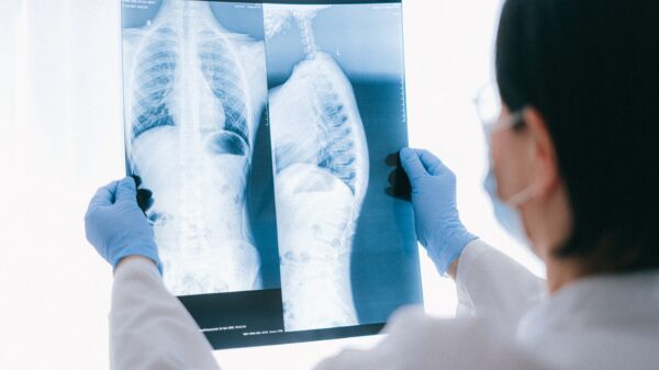
Breast cancer is the most common diagnosed cancer among women
The research team believes the dual-mode imaging system could eventually monitor patients throughout treatment. Consequently, doctors may gain earlier insight into whether therapies are shrinking tumours or failing to respond.
According to the Canadian Cancer Society, breast cancer remains the most commonly diagnosed cancer among Canadian women. About one-in-eight women in Canada are expected to receive a breast cancer diagnosis during their lifetime.
LoVetri said nearly every Canadian family knows someone affected by the disease. Additionally, he believes earlier detection and improved monitoring could help reduce long-term impacts for patients.
The project has now entered a new phase focused on commercialization and human testing. The University of Manitoba has partnered with Taumedis Inc., a Winnipeg-based medical imaging company, to guide the technology toward clinical use.
Taumedis co-founder Michael Lang said the company will help navigate regulatory approvals, testing requirements and future clinical trials. Furthermore, the partnership aims to bridge the gap between university research and a commercially viable medical product.
Lang said several hurdles remain before the technology reaches hospitals or clinics. Human studies, ethics approvals and regulatory reviews must all proceed before broader adoption becomes possible.
The first human study involving the imaging system is scheduled for later this year, pending regulatory and ethics approval. Additionally, researchers expect those trials to provide critical data on patient comfort and imaging performance.
Lang said the timeline could shift as development continues. However, the upcoming studies should provide researchers with a clearer understanding of how the technology performs in real-world conditions.
Read more: Breath Diagnostics advances pre-op pneumonia screening with FDA breakthrough designation
Read more: Breath Diagnostics leaders promote their mission at Miami investment conference
Work reflects a broader push for cancer detection
Master’s student Skylar Trudeau described the project as a rare opportunity to work on a potentially groundbreaking medical technology. He said few research groups worldwide currently combine ultrasound and microwave imaging in the same system.
Trudeau also noted that many women find mammograms uncomfortable. Consequently, researchers hope the dual-mode system could eventually provide another option alongside traditional screening tools.
The work at the University of Manitoba reflects a broader push to improve how doctors detect and monitor cancer through less invasive imaging systems. Additionally, researchers worldwide are developing technologies that combine artificial intelligence, advanced sensors and new scanning methods to identify tumours earlier and track treatments more accurately.
Many existing screening methods still face significant limitations. Mammograms can produce inconclusive results, while some cancers remain difficult to identify until symptoms appear. Several screening procedures remain uncomfortable or invasive, discouraging some patients from seeking early testing.
As a result, companies and research groups have shifted toward earlier, simpler and more patient-friendly diagnostic tools. Grail Inc (NASDAQ: GRAL), for example, developed the Galleri blood test to identify signals linked to multiple cancer types from a single blood sample. The company says the technology can help detect cancers that currently lack routine screening options.
Meanwhile, Breath Diagnostics Inc. has been developing its OneBreath technology for lung cancer screening. The system analyzes compounds in a patient’s breath that may indicate the presence of disease. Researchers hope the approach could offer a fast and non-invasive method for identifying lung cancer earlier.
.