
University of Southern California researchers are developing quantum-enhanced artificial intelligence that could help doctors identify and treat cancer more precisely.
Announced on Tuesday, the researchers created a hybrid quantum-classical system designed to improve how artificial intelligence analyzes medical images. Their approach could eventually help doctors map tumors more accurately while reducing the computing power needed for the task.
Amir Kalev, lead quantum scientist at USC Viterbi’s Information Sciences Institute, leads the research. He also serves as an adjunct research professor in USC’s Department of Physics and Astronomy.
Kalev has focused his quantum computing research on healthcare because medical imaging requires highly precise predictions. Additionally, mistakes in interpreting those images can directly affect how doctors diagnose and treat patients.
Doctors regularly use magnetic resonance imaging, computed tomography and ultrasound scans to diagnose diseases and plan treatments. Increasingly, artificial intelligence systems help physicians interpret the enormous amounts of information contained in those images.
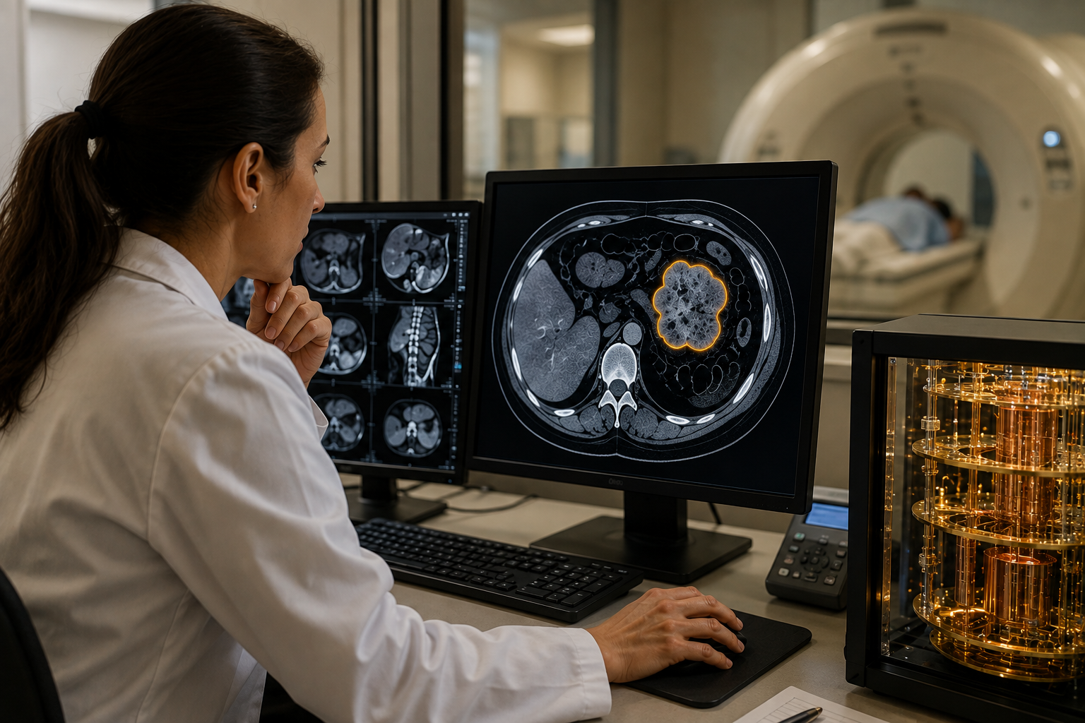
However, those systems must first identify exactly where abnormal tissue begins and healthy tissue ends. Researchers call this process image segmentation.
Image segmentation essentially teaches a computer to trace the boundaries of an object inside an image. In cancer care, the object could be a tumor surrounded by healthy organs and other tissue.
The system examines individual pixels and determines which belong to the tumor. Consequently, more accurate segmentation can provide doctors with a clearer picture of a tumor’s shape, size and location.
Those boundaries can affect several stages of cancer treatment. Doctors can use them to plan surgery, direct radiation and track whether tumors respond to therapy.
Read more: Prestigious medtech intelligence firm recognizes Breath Diagnostics for innovation
Read more: Breath Diagnostics completes install of advanced mass spectrometry system
QuFeX designed to strengthen existing AI systems
Additionally, better boundaries could help physicians avoid damaging nearby healthy organs during treatment. That could improve outcomes while reducing unwanted side effects for patients.
Kalev suspected quantum computing could improve the ability of artificial intelligence to draw those boundaries. He worked with former student Naman Jain to develop a quantum module called Quantum Feature Extraction, or QuFeX.
Jain earned a master’s degree in quantum information science in 2025. Together, the researchers designed QuFeX to strengthen existing artificial intelligence systems rather than replace conventional computing.
Their research appeared in the journal Quantum Science and Technology. Furthermore, the researchers built the project around a significant problem facing medical artificial intelligence: limited data.
Large artificial intelligence models often improve when developers train them on enormous datasets. Medical researchers do not always have that luxury because high-quality medical datasets can remain relatively small.
Patient privacy, specialized imaging requirements and limited examples of certain diseases can constrain available training information. In addition, medical images can vary substantially in quality and consistency.
Jain said the team wanted to determine whether quantum technology could help artificial intelligence overcome those limitations. The researchers subsequently incorporated QuFeX into an established medical-imaging system called U-Net.
U-Net is a neural network architecture widely used for image segmentation. The resulting hybrid system, called Qu-Net, combines conventional artificial intelligence with quantum computing techniques.
The researchers then compared Qu-Net with a leading conventional artificial intelligence model across several image-segmentation benchmarks. Those tests included datasets involving medical images.
Read more: Breath Diagnostics advances pre-op pneumonia screening with FDA breakthrough designation
Read more: Breath Diagnostics leaders promote their mission at Miami investment conference
Quantum computing’s value depends on efficiency
Qu-Net beat the classical baseline by roughly 7 per cent, according to Kalev. Meanwhile, the hybrid model achieved that result with substantially fewer adjustable parameters.
Parameters are internal values that an artificial intelligence system learns during training. They help determine how the system interprets information and produces its predictions.
Qu-Net used approximately 250,000 parameters during the researchers’ tests. Conversely, U-Net required approximately 1.5 million parameters, or about six times as many.
That difference could become important if quantum-enhanced systems eventually move into hospitals. Smaller models can require less computing power and potentially consume fewer resources during training.
Additionally, fewer parameters can make sophisticated artificial intelligence tools easier to deploy where computing resources remain limited. The result could matter beyond raw processing speed.
Jain said quantum computing’s value may depend partly on efficiency rather than simply outperforming conventional machines in speed. Furthermore, Qu-Net produced more accurate boundaries around suspicious tissue during the researchers’ experiments.
Those improvements could eventually help radiation oncologists determine which areas require treatment. Doctors must carefully target tumors while limiting radiation exposure to surrounding healthy tissue.
However, the research remains at an early stage and has not yet established Qu-Net as a clinical diagnostic tool. Kalev has started working with physicians at USC’s Keck School of Medicine to investigate practical applications.
The collaboration includes Dr. Eric Chang, chair of Keck’s Department of Radiation Oncology. Together, the researchers are examining whether quantum machine learning can improve radiation-treatment planning.
Radiation oncologists currently spend significant time outlining tumors and nearby organs before delivering treatment. Additionally, specialists may perform some of that segmentation manually to ensure treatment targets the correct areas.
Read more: Breath Diagnostics leader speaks at lung cancer education event in Louisville
Read more: Breath Diagnostics now offering a compelling investment opportunity
Technologies are adjustable for changing circumstances
Chang sees an opportunity to reduce that workload through more capable image-analysis systems. Automating parts of segmentation could allow medical teams to prepare radiation plans more efficiently.
The researchers plan to test their technology using simulated scans and images from real patients. Subsequently, successful testing could lead to systems that help physicians adjust radiation plans as tumors change.
Tumors can shrink, grow or change shape during treatment. Meanwhile, surrounding organs and other tissues can also move or change between treatment sessions.
Those changes can require doctors to revise a patient’s radiation plan. A faster segmentation system could potentially help physicians make those adjustments without lengthy delays.
Kalev ultimately wants to shorten personalized radiation-treatment planning from days to a single clinical visit. Under that approach, doctors could scan a patient, develop the plan and potentially begin treatment much sooner.
Furthermore, faster planning could reduce the time patients spend waiting between diagnosis and treatment. The technology would still require extensive testing before researchers could establish whether it delivers those benefits in clinical practice.
The team also sees possible applications outside cancer care. Similar image-analysis techniques could eventually assist doctors working with brain disorders, cardiovascular disease and surgical planning.
In addition, the underlying technology could have applications outside medicine wherever computers must interpret complicated images. Possible uses include autonomous vehicles, satellite imagery and other computer-vision systems.
Kalev views QuFeX and Qu-Net primarily as attempts to turn quantum research into practical tools. Rather than proving quantum computing’s value through theoretical performance alone, his team aims to address problems that directly affect patients.
Read more: Breath Diagnostics adopts state-of-the-art mass spectrometer device for lung screening
Read more: Breath Diagnostics tech achieves pneumonia prediction breakthrough in peer-reviewed study
New technologies enter the fight against cancer
Quantum computing represents only one of several technologies researchers are bringing into the fight against cancer. Scientists are also turning to artificial intelligence, liquid biopsies and increasingly unconventional methods to detect the disease earlier.
Artificial intelligence already helps analyze mammograms, CT scans and other medical images for suspicious tissue. In March, U.S. regulators approved an AI-assisted imaging system from Perimeter Medical Imaging AI Inc. (CVE: PINK) (OTCMKTS: PYNKF). Its Claire system helps surgeons identify areas suspicious for breast cancer while examining tissue removed during lumpectomies.
Meanwhile, researchers are investigating whether cancer can reveal itself through something as simple as a patient’s breath. Breath Diagnostics has developed OneBreath, which analyzes volatile organic compounds exhaled from the lungs. The company says its system uses mass spectrometry and AI to identify molecular signatures associated with lung cancer.
Other researchers have taken the idea of smelling cancer considerably further.
Scientists have trained dogs to identify chemical signatures associated with cancer in breath samples. A 2026 study involving 3,275 participants tested trained dogs across seven major cancer groups at six hospitals in India. Researchers combined the animals’ responses with mathematical modelling to produce cancer-risk assessments.
Researchers have even experimented with microscopic worms. Studies involving the nematode Caenorhabditis elegans found that the worms respond differently to urine depending on whether cancer-related chemical signals are present.