Researchers in Quebec have developed a new imaging system that can detect melanoma days before it becomes visible, potentially giving doctors a powerful new tool to identify aggressive skin cancers at their earliest stages.
Late last month, scientists from the Institut national de la recherche scientifique and Université de Montréal tested the technology in mice and found it could identify tiny melanoma tumours just four days after they formed. The team recently published its findings in the journal Nature Sensors.
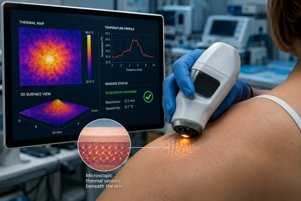
Called SMEAR-ULM, the system measures subtle temperature differences on the skin’s surface. Additionally, it converts those thermal signals into detailed maps that can reveal the presence of cancer long before traditional methods can detect it.
Melanoma rates continue to rise across Canada. Meanwhile, doctors still rely heavily on visual inspections followed by biopsies to confirm whether suspicious skin lesions are cancerous. Those procedures can be invasive and may sometimes prove unnecessary.
The researchers designed SMEAR-ULM to offer a faster and less invasive alternative. Consequently, clinicians could eventually use the technology to assess questionable lesions before deciding whether a biopsy is needed.
The project brought together several research groups. INRS professor Jinyang Liang led the work alongside collaborators including INRS professor Fiorenzo Vetrone, Université de Montréal pharmacology professor Davide Brambilla and medical professor Sylvain Meloche.
Liang said the team wanted to create a tool capable of identifying very small but potentially dangerous melanomas. Furthermore, he explained that doctors often exclude lesions of that size from routine visual examinations because they remain difficult to see. Early detection could allow treatment to begin sooner.
Read more: Prestigious medtech intelligence firm recognizes Breath Diagnostics for innovation
Read more: Breath Diagnostics completes install of advanced mass spectrometry system
Nanoparticles act as microsocope thermometers
The technology takes a different approach from conventional cancer imaging systems. Researchers have long known that tumours generate extra heat because cancer cells consume more energy than healthy tissue. However, doctors have struggled to use that heat as a reliable diagnostic signal because existing thermal imaging systems lack sufficient precision.
At the centre of SMEAR-ULM sits a patch containing painless microneedles. The patch delivers specialized nanoparticles just beneath the skin, creating what researchers describe as a temporary intelligent tattoo.
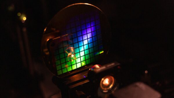
Those nanoparticles act as microscopic thermometers. When researchers illuminate them with near-infrared light, they emit visible light in response. Additionally, the duration of that light emission changes according to local temperature conditions.
Cancer cells typically consume more oxygen and nutrients than surrounding healthy tissue. Consequently, they produce extra heat that alters the nanoparticles’ optical behaviour. Researchers can then measure those changes to identify abnormal tissue.
The system pairs those nanoparticles with an ultrafast imaging platform capable of capturing all relevant information in a single snapshot. Furthermore, it produces highly detailed temperature maps with submillimetre spatial resolution and sensitivity to temperature changes smaller than one degree.
First author Yingming Lai, an INRS postdoctoral fellow, said the technology generates an instantaneous temperature map from a single image. As a result, researchers can monitor unusual thermal activity in tiny melanomas quickly and reliably, even under complex living conditions.
Read more: Breath Diagnostics advances pre-op pneumonia screening with FDA breakthrough designation
Read more: Breath Diagnostics tech achieves pneumonia prediction breakthrough in peer-reviewed study
Microneedle-based sensing technologies require repeated measurements
During testing, the researchers detected micro-melanomas only four days after they appeared. Conversely, conventional thermal imaging systems generally identify tumours only after they exceed five millimetres in size, making them visible to the naked eye.
Most existing thermal imaging systems rely on infrared technology. However, those systems often suffer from limited resolution and significant background noise, reducing their ability to detect extremely small lesions.
Researchers also noted that other microneedle-based sensing technologies often require repeated measurements. Meanwhile, SMEAR-ULM gathers its data in real time through a single-shot imaging process.
Meloche said the mouse model used in the study closely replicates genetic changes found in human melanoma. Additionally, he noted that the findings suggest the technology could eventually provide benefits for human patients if future studies confirm its effectiveness.
Beyond melanoma detection, the researchers believe the platform could support other biomedical applications. Furthermore, they said future versions could measure factors such as pH levels or ion concentrations, potentially expanding its role in medical imaging and diagnostic research.
The concept reflects a broader shift toward biomarker-driven diagnostics. Breath Diagnostics has pursued a similar objective through breath-analysis technology designed to detect disease-related chemical signatures. Rather than relying solely on visible symptoms, these approaches attempt to identify measurable biological changes associated with disease.
.