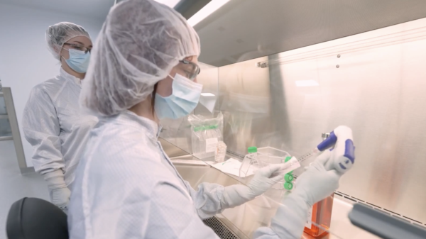
A groundbreaking method developed by researchers at the Fraunhofer Institute could significantly reduce radiation exposure during cancer diagnosis and treatment.
Announced on Tuesday, this new system combines traditional X-ray imaging with radar technology. Further, it offers a safer, more accurate alternative for detecting and monitoring breast and lung cancer.
X-ray mammography remains the leading tool in early breast cancer detection. It provides fast and precise two-dimensional images. Computed tomography (CT) adds valuable three-dimensional imaging for lung cancer diagnosis. However, CT scans deliver high radiation doses. While natural annual radiation exposure is about 2.1 millisieverts, a single chest CT scan can triple that amount.
The Fraunhofer-led MultiMed project aims to solve this challenge by merging radar and X-ray imaging into one system. This multimodal imaging method could transform medical diagnostics and significantly reduce risks to patients.
Radar technology has long played a vital role in aviation and automotive industries. However, its role in medicine has been limited until now. Additionally, radar offers three-dimensional imaging capabilities without the harmful side effects of ionizing radiation. Therefore, integrating radar into medical diagnostics opens new opportunities for safer cancer detection.
Radar cannot match the resolution or depth of conventional imaging. However, it detects changes in tissue based on differences in electrical permittivity and conductivity. This unique property helps identify abnormal tissue growth. In addition, radar can provide material information that traditional imaging methods cannot offer directly.
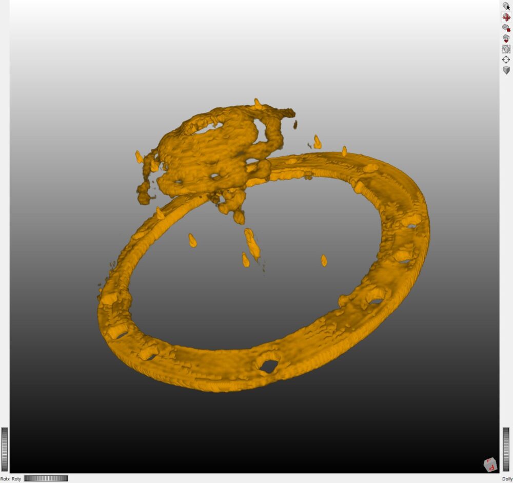
The core challenge lies in uniting data from radar and X-rays into a single coherent image. To address this, researchers are developing a process called “co-registering.” This method spatially aligns radar and X-ray data, allowing the images to complement one another.
Read more: Breath Diagnostics gives the public the chance to join the fight against cancer
Read more: Breath Diagnostics leader speaks at lung cancer education event in Louisville
Hybrid technique boosts clarity
Additionally, advanced radar reconstruction algorithms are in development. These will sharpen radar images and enhance tissue differentiation.
Further improvements are underway in CT imaging as well. By incorporating radar data into CT image reconstruction, the research team is creating a multimodal CT algorithm. This hybrid technique boosts image clarity, reduces visual artifacts, and lowers radiation doses. Therefore, patients may benefit from a more comfortable and less risky diagnostic process.
Researchers have already built imaging phantoms to test the new approach. These phantoms mimic human tissue and generate accurate signals for both radar and X-ray systems. Accordingly, they serve as valuable tools for validating the combined imaging method in laboratory settings.
In addition, the project’s goal is to produce a fully functional lab system by the end of the three-year development phase. This system will integrate CT and radar imaging into one platform. Furthermore, it will serve as a test bed for medical applications and further research.
“The new approach has the potential to detect tissue changes early on and with great accuracy—and with much less of a burden on patients than before,” says Victoria Heusinger-Hess, the project manager at the Fraunhofer Institute for High-Speed Dynamics, Ernst-Mach-Institut (EMI).
This development comes at a crucial time. Cancer remains a leading cause of death worldwide, and the risks associated with repeated imaging must be managed. Accordingly, reducing radiation exposure without compromising diagnostic accuracy is vital.
The fusion of radar and X-ray imaging presents a unique step forward in this effort. It also sets the stage for future medical imaging systems that are safer and more informative. Additionally, the ability to detect subtle tissue changes early could improve treatment outcomes for millions.
Read more: Breath Diagnostics opens Respiratory Innovation Summit with captivating presentation
Read more: Breath Diagnostics now offering a compelling investment opportunity
Chest CT exposes patients to over three times annual radiation exposure
X-ray technology plays a vital role in modern diagnostics, but it carries health risks that cannot be ignored. Exposure to ionizing radiation can damage DNA and increase cancer risk over time. A standard chest X-ray emits a small dose of radiation, but CT scans—especially full-dose—deliver far more. A chest CT can expose a patient to as much as 7 millisieverts of radiation, or over three times the annual natural background exposure.
To reduce this risk, the medical community has adopted low-dose CT as a standard of care in many situations, particularly for lung cancer screening. This approach cuts radiation by using fewer X-ray beams or lower energy levels. However, even low-dose CT involves radiation. When used repeatedly, especially in long-term monitoring, the cumulative effect can still be significant.
Accordingly, researchers and companies are working to create diagnostic alternatives that eliminate radiation exposure entirely. These technologies aim to detect disease through physical or chemical signals, rather than imaging alone. One such company is Breath Diagnostics, based in Kentucky. It focuses on analyzing exhaled breath to identify volatile organic compounds linked to cancers such as lung and breast.
Furthermore, breath-based diagnostics offer speed, comfort, and repeatability. They can be administered frequently without any health risk. Additionally, these tools are non-invasive and well-suited for both early screening and ongoing monitoring.
While traditional imaging remains essential, tools like those developed by Breath Diagnostics show that safer, radiation-free methods are within reach. These innovations could soon complement or, in some cases, replace standard scans. Therefore, the future of diagnostics may be not only more accurate, but also far gentler on the human body.
.
joseph@mugglehead.com

