What if a tattoo, a nanoparticle, or even your breath could reveal cancer before it strikes? Scientists are turning fiction into life-saving reality.
Lung cancer kills more people than any other cancer, yet according to the American Cancer Society only about 6 per cent of eligible Americans undergo annual screening.
Low-dose computed tomography (LDCT) scans can reduce lung cancer deaths by up to 20 per cent, but only if patients actually show up—and keep coming back. Adherence drops from 61 per cent in the first year to just over 50 per cent by year two.
Part of the problem lies in the scans themselves. LDCT uses low doses of radiation to detect tumours early. Ironically, repeated exposure can slightly increase cancer risk, creating a paradox where screening to prevent cancer may also contribute to it.
This dilemma highlights the urgent need for safer, more effective alternatives. Researchers are exploring innovative ways to detect and treat cancer without relying on radiation alone. From spider silk delivering chemotherapy directly to tumours, to smart tattoos monitoring tumour progression in real time, science is inventing tools that are precise, minimally invasive, and patient-friendly.
These approaches also offer earlier detection for people who fall outside traditional screening criteria, such as non-smokers or those exposed to environmental carcinogens. Furthermore, reducing reliance on invasive procedures or repeated scans may improve compliance and overall outcomes.
Meanwhile, clinicians and patients are watching closely. The future of oncology could combine safety, speed, and precision, catching tumours earlier and treating them more effectively—without the hidden risks of low-dose radiation.
1. Fluorescent bacteria will help find tumours in the future
Bacteria that light up tumours are revolutionizing cancer surgery. Scientists have engineered bacteria that activate specifically within tumour tissue, allowing surgeons to locate tumours in real time. This method enables more precise tumour removal and reduces the risk of recurrence.
The bacteria emit fluorescent light that shows tumour margins like a neon sign. Consequently, surgeons can distinguish cancerous tissue from healthy cells more easily. Furthermore, the fluorescence remains visible for days, even in complex internal organs, giving doctors a longer window for accurate imaging.
This approach was developed by a team led by Dr. SeungBeum Suh at the Korea Institute of Science and Technology, in collaboration with Professor Hyo-Jin Lee from Chungnam National University Hospital. Their work focuses on improving surgical precision and patient outcomes by providing real-time tumour visualization.
The bacteria accumulate in tumours because cancer cells create a unique environment that normal tissue cannot support. Subsequently, healthy tissue remains mostly unaffected, minimizing side effects. In addition, this targeted approach could complement existing imaging techniques, offering a dynamic view of tumour progression.
Researchers are also exploring ways to combine glowing bacteria with treatments that attack cancer cells directly. Meanwhile, improvements in imaging technology enhance the sensitivity of tumour detection, making this method even more effective.
While still experimental, bacteria that light up tumours represent a major step forward in oncology. By turning living cells into diagnostic tools, scientists hope to catch cancers earlier, improve surgical outcomes, and reduce unnecessary treatments. This technology could transform how doctors find and treat tumours, offering patients a brighter prognosis.
Read more: Breath Diagnostics leader speaks at lung cancer education event in Louisville
Read more: Breath Diagnostics opens Respiratory Innovation Summit with captivating presentation
Spider silk delivers chemotherapy
Spider silk, known for its remarkable strength and flexibility, is now showing promise as a vehicle for cancer treatment. Researchers have found that the protein fibers can carry chemotherapy drugs directly to tumours while sparing healthy tissue. Its biocompatibility and biodegradability make spider silk an ideal delivery system for medicine.
Chemotherapy can harm healthy cells while attacking cancer. This causes side effects like nausea, fatigue, and hair loss. Researchers hope spider silk nanoparticles can solve that problem. The silk fibers can encapsulate drugs and release them slowly, targeting only cancer cells. This approach reduces the impact on healthy tissue.
In laboratory studies, scientists loaded chemotherapy agents into tiny silk-based carriers. They observed that the carriers reached tumours efficiently and released the drugs in a controlled way. Furthermore, the silk broke down naturally in the body after delivering the treatment. Consequently, the therapy did not leave harmful residues.
This method also allows researchers to combine multiple drugs in a single carrier. In addition, they can adjust the size, shape, and composition of the silk particles to match different tumor types. The flexibility of the system makes it highly adaptable for personalized cancer treatment.
Researchers are now moving toward animal studies and early clinical trials. They aim to confirm safety and effectiveness in living organisms. Meanwhile, pharmaceutical companies are exploring partnerships to scale production.
If successful, spider silk drug delivery could change chemotherapy forever. By providing precise, targeted therapy, this approach promises fewer side effects and better outcomes. Furthermore, it opens the door to using other natural materials in advanced cancer treatment, demonstrating how biology can inspire innovation in medicine.
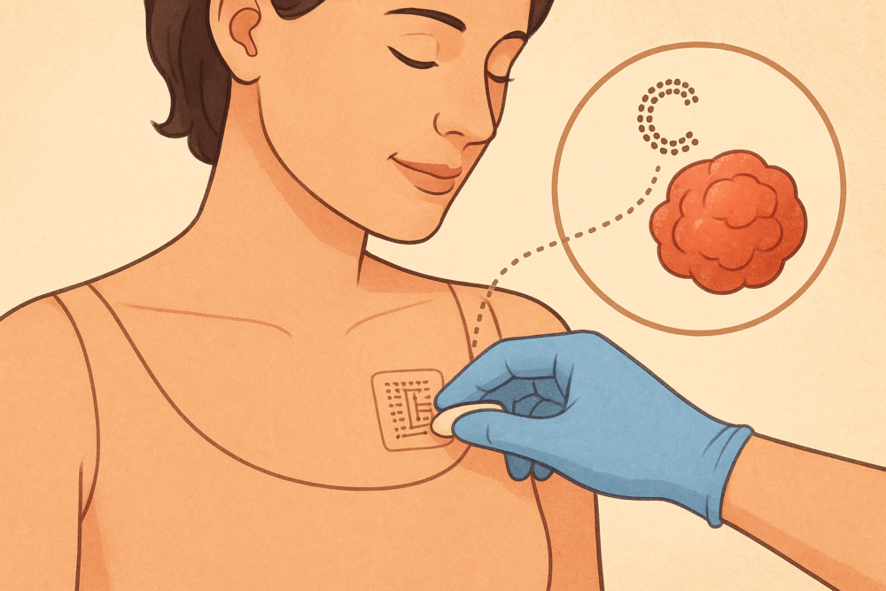
Smart tattoos monitor tumor progression
More than just a way to piss your parents off, scientists have developed smart tattoos that can monitor cancer from inside the body.
These tiny, flexible sensors are implanted under the skin and can track tumour growth or treatment response in real time. The tattoos change colour or transmit data when biological markers such as pH or oxygen levels shift, which often signals tumour activity.
Researchers from the Swiss Federal Institute of Technology (ETH Zurich) first demonstrated the concept, showing that smart tattoos could detect calcium changes linked to tumour growth. Since then, several teams across Europe, the United States, and Asia have expanded on the idea, creating bioresorbable sensors that safely dissolve after use. These new materials eliminate the need for surgical removal and make long-term monitoring safer for patients.
The technology combines biochemistry and microelectronics. Sensors respond to chemical changes inside the body and send data wirelessly to an external device. In addition, doctors can use the readings to assess whether a tumour is shrinking or becoming resistant to therapy. Consequently, treatment can be adjusted quickly, improving the chances of success.
Furthermore, smart tattoos are being designed to track other diseases, such as diabetes or cardiovascular disorders. Their ability to provide continuous data without invasive procedures represents a major shift in how medicine is practiced.
Although the technology remains in early testing, clinical trials are expected within the next few years. If successful, smart tattoos could give oncologists a clear, continuous view of tumor behavior, reducing the need for repeated biopsies.
Read more: Breath Diagnostics leaders promote their mission at Miami investment conference
Read more: Breath Diagnostics gives the public the chance to join the fight against cancer
Magnetic nanoparticles target and destroy tumors
Scientists are using magnetic nanoparticles to destroy tumors from the inside out. The technology, known as magnetic hyperthermia, relies on tiny iron oxide particles that heat up when exposed to an alternating magnetic field. Once injected into a tumor, these nanoparticles generate localized heat that kills cancer cells while leaving surrounding tissue unharmed.
Researchers from institutions such as the Max Planck Institute for Intelligent Systems in Germany, Rice University in the United States, and the National Cancer Institute are leading the development. The technique has already shown success in preclinical studies and small human trials, especially for brain, prostate, and breast cancers.
The process begins when doctors inject the nanoparticles directly into or near the tumor site. After that, an external magnetic field is applied. The particles vibrate and release controlled heat, reaching about 42 to 45 degrees Celsius — just enough to damage or destroy cancer cells without harming healthy ones. Furthermore, the heat can make tumors more sensitive to chemotherapy or radiation, improving treatment effectiveness.
In addition, researchers are coating the nanoparticles with antibodies or polymers that help them bind only to cancer cells. This targeted delivery increases safety and precision. Some teams are even designing particles that can carry drugs, combining hyperthermia and chemotherapy in a single treatment.
Clinical studies in Europe and Asia have demonstrated promising results, showing tumor shrinkage and longer survival in certain patients. However, scientists continue to refine the method to ensure even heat distribution and minimize side effects.
Consequently, magnetic nanoparticle therapy represents a major shift in cancer treatment. By using magnetism instead of scalpels or radiation, it offers a minimally invasive, highly targeted way to fight cancer — one that could redefine how tumors are treated in the years ahead.
Breath Diagnostics detects lung cancer via exhaled biomarkers
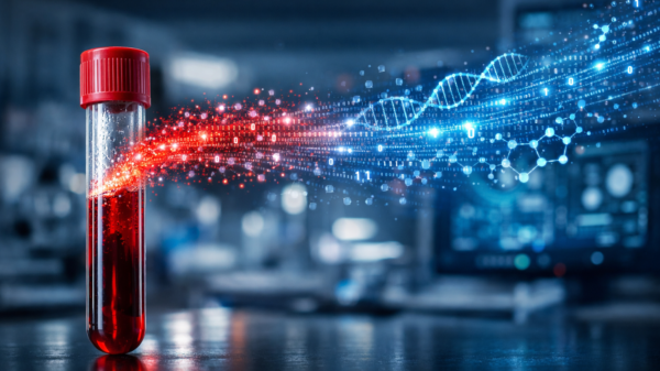
Breath Diagnostics Inc., a Kentucky-based startup founded in 2015, is pioneering a non-invasive approach to detecting lung cancer. The company developed OneBreath, a platform that analyzes volatile organic compounds (VOCs) in a single exhaled breath. These compounds act as biomarkers, revealing early signs of disease. A proprietary microreactor chemically transforms the VOCs, which are then measured using ultra-high-performance liquid chromatography-mass spectrometry (UHPLC-MS). This process captures even trace compounds without radiation or invasive procedures.
OneBreath™ has shown strong clinical validation, with a sensitivity of 94 per cent and specificity of 85 per cent across multiple independent sites and stages of disease. Its AI-powered analysis interprets complex VOC patterns, enabling early detection of lung cancer, respiratory infections, and gastrointestinal disorders. By harnessing the natural process of breathing, the platform offers a faster and safer alternative to traditional methods like CT scans or biopsies.
The company is now expanding trials to include a broader range of patients across North America. In addition, researchers are studying how lifestyle, diet, and environmental exposures influence VOC profiles. Early results suggest OneBreath can detect lung cancer in people who do not smoke, a group often missed by conventional screening. Furthermore, the platform provides rapid results, often within minutes, improving patient outcomes.
Breath Diagnostics is also engaging in a regulation crowdfunding offering to raise additional capital. By combining non-invasive testing with AI-driven interpretation, the company aims to scale its technology and reach more patients, potentially improving early detection and reducing mortality from lung cancer.
.
joseph@mugglehead.com